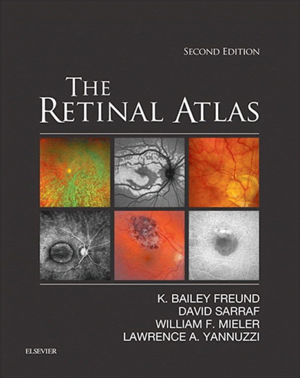
کتاب The Retinal Atlas | اطلس شبکیه چشم ویراست دوم 2017 با بیش از 5000 تصویر، یک صفحه آرایی منحصر به فرد ، و تصاویر جامع از کل طیف اختلالات زجاجیه ، شبکیه و ماکولا ، اطلس شبکیه مرجع ضروری برای متخصصان شبکیه چشم و چشم پزشکان جامع و سایر علاقمندان است.
With more than 5,000 images, a unique page layout, and comprehensive illustrations of the entire spectrum of vitreous, retina, and macula disorders, The Retinal Atlas, 2nd Edition, is an indispensable reference for retina specialists and comprehensive ophthalmologists as well as residents and fellows in training. For this edition, an expanded author team made up of Drs. K. Bailey Freund, David Sarraf, William F. Mieler, and Lawrence A. Yannuzzi, each an expert in retinal research and imaging, provide definitive up-to-date perspectives in this rapidly advancing field.
This award-winning title has been thoroughly updated with new images with multimodal illustrations, new coverage and insight into key topics, and new disorders and classifications, while retaining the innovative page layout that has made it the most useful and most complete atlas of its kind.
- Provides a complete visual guide to advanced retinal imaging and diagnosis of the full spectrum of retinal diseases, including early and later stages of disease.
- Enhances understanding by presenting comparison imaging modalities, composite layouts, high-power views, panoramic disease visuals, and selected magnified areas to hone in on key findings and disease patterns.
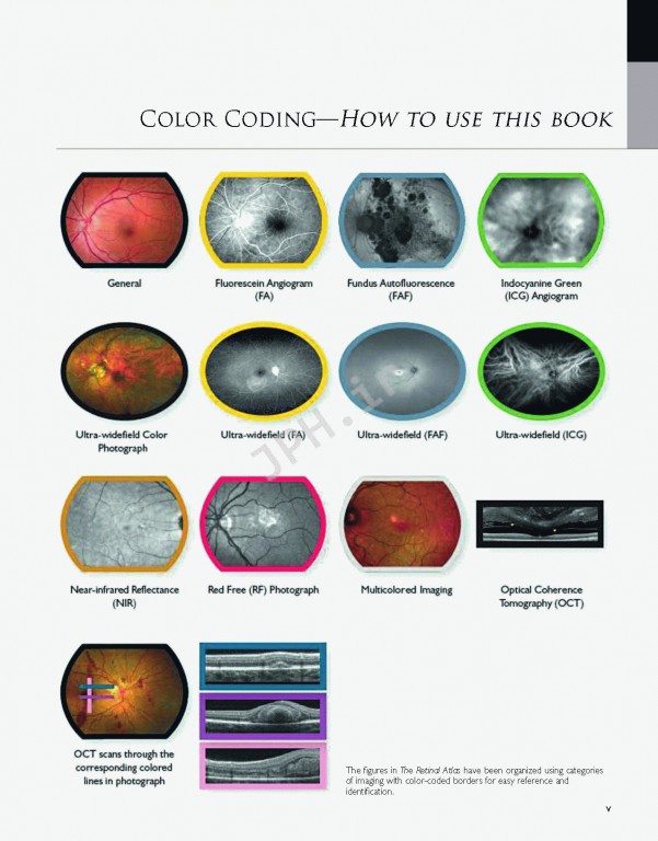
- Features color coding for different imaging techniques, as well as user-friendly arrows, labels, and magnified images that point to key lesions and intricacies.
- Expert Consult™ eBook version included with purchase. This enhanced eBook experience allows you to search all of the text, figures, and references from the book on a variety of devices.
- Covers all current retinal imaging methods including: optical coherence tomography (OCT), indocyanine green angiography, fluorescein angiography, and fundus autofluorescence.
- Depicts and explains expanding OCT uses, including spectral domain and en face OCT, and evolving retinal imaging modalities such as ultra-wide-field fundus photography, angiography and autofluorescence.
- Presents a select team of experts, all of whom are true international leaders in retinal imaging, and have assisted in contributing to the diverse library of common and rare case illustrations.